X Ray Mastoid Laws View

X Ray Mastoid Lateral Oblique Law S View Left Side Shows Sclerosis Download Scientific Diagram

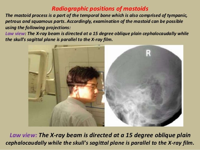
Radiographic Positions Of Mastoids Human Head And Neck Human Anatomy
Radiology Quiz Radiopaedia Org
Pubs Rsna Org Doi Pdf 10 1148 80 2 255

Cochlear Implant Otorhinolaryngology Portal
Cone Beam Ct In Dental Practice British Dental Journal
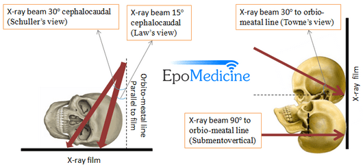
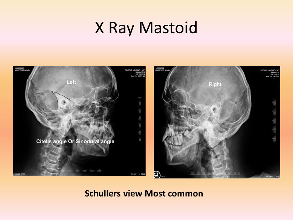
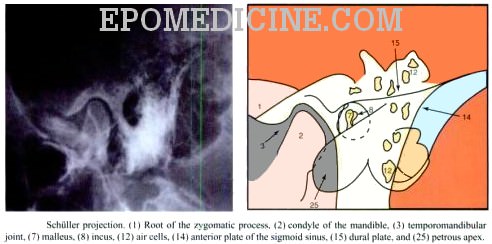
The Schullers view serves as an alternative view to the Law projection which uses a 15 degree angle of patient's face toward the image receptor and a 15 degree caudal angulation of the CR to achieve the same result, a lateral mastoid air cells view without overlap of.
X ray mastoid laws view. Skull, a-p, tilted X-ray, child 5 months. This is a normal mastoid series for reference. You Can learn the easiest X-Ray of Mastoid Townes View From this Video.
The X-ray beam is directed at a 15 degree oblique plain cephalocaudally while the skull’s sagittal plane is parallel to the X-ray film. X-ray of mastoids, lateral and oblique views. It is my own practice at the present time at Gray's Inn Road and.
You are required to remain still during the process of scanning. Know why the test is suggested, how to prepare, benefits, risks and more. Pterion (sphenoidal fontanelle) Greater wing of.
X-ray both mastoids Laws view (lateral oblique) Differential diagnosis:. The waves scan the internal condition of the area and produce an image onto the computer screen. The x-ray study of the mastoid region, which was begun in March, 1908, has undergone a slow but gratifying metamorphosis.Undertaken with grave doubts as its practical value, it has developed into a method which rivals in its accuracy other recognized methods of physical examination.
"I always getting headache pain behind my ear around my back neck and forehead my BP is 129/94. A plain X-ray of mastoid/Law’s view was done to assess the position of dural and sinus plates. For the best hearing outcomes, a minimum of 15 intra-cochlear electrodes is required 3.
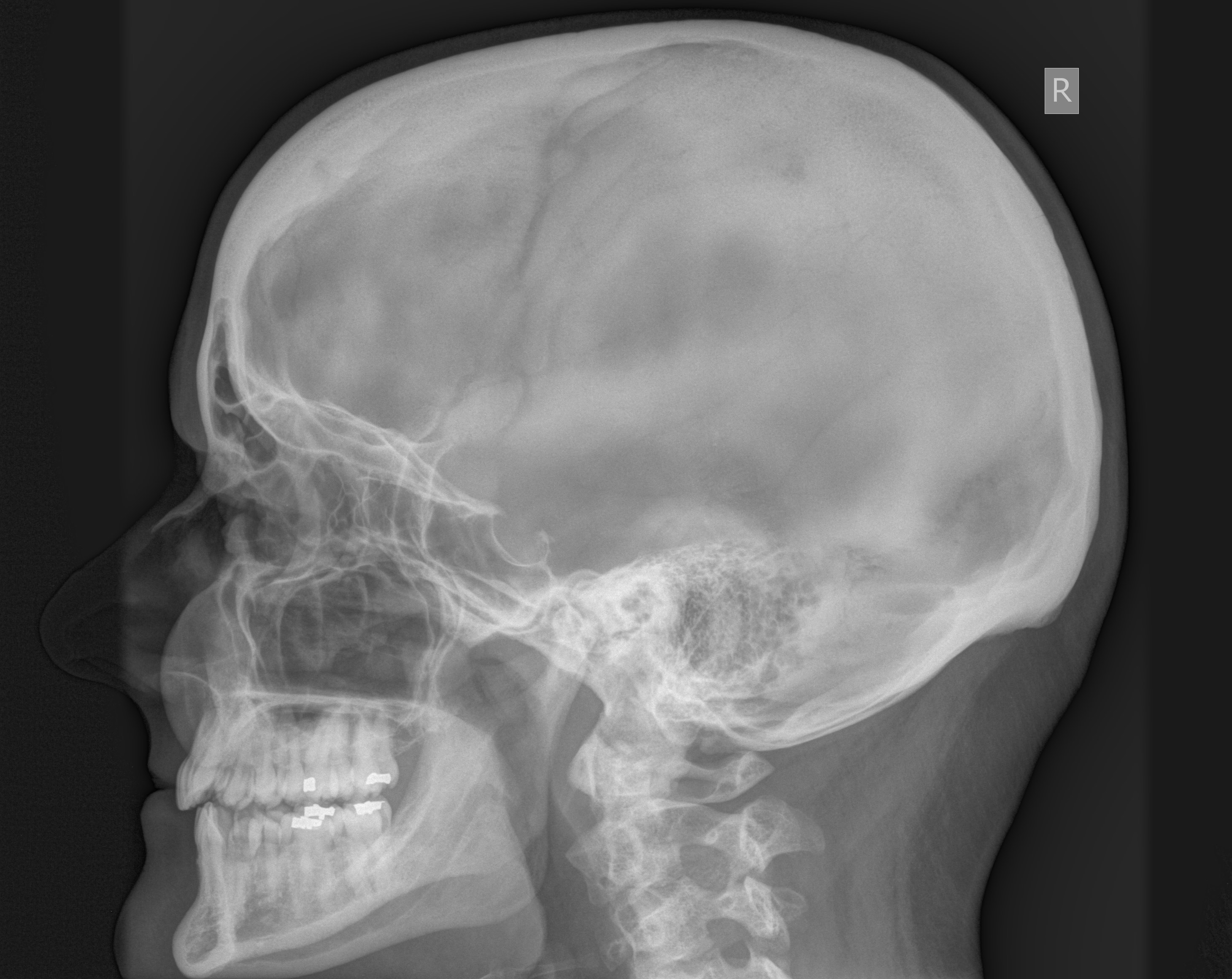
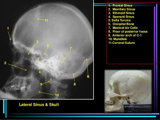
Chest X-ray (Each Oblique View) Orbit X. This is an x-ray image of the skull taken from a lateral view showing the skull from the side. X-ray of mastoid, less than 3 views per side.
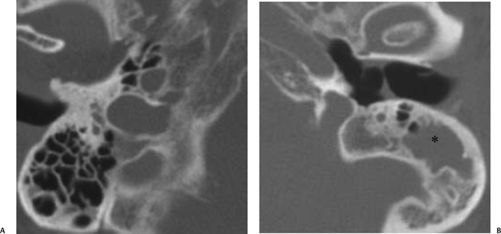
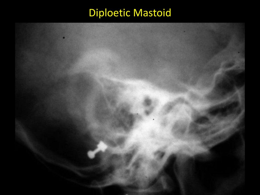
Become a single cavity seperated by middle ear cavity. Large antral cell - This is usually bilateral. Cholesteatomatous cavity - Radiologically this cavity will be surrounded by a rim of sclerosis.
Usually these projection taken in open and closed mouth positions. Axiolateral Oblique-Modified Law Method- CENTRAL RAY -CR directed to a midpoint of the grid at an angle of 15 degrees caudad to exit the downside mastoid tip approximately 1 inch posterior to the EAM -The CR enters approximately 2 inches posterior to, and 2 inches superior to the uppermost EAM. X-ray femur 2 views x-ray knee 1-2 views x-ray knee 3 views x-ray knee 4+ views x-ray bilateral knees standing x-ray tibia fibula 2 views x-ray ankle 2 views x-ray ankle 3+ views 736 x-ray foot, two views x-ray foot, 3+ views x-ray heel 2+ views x-ray toe--2 or more views.
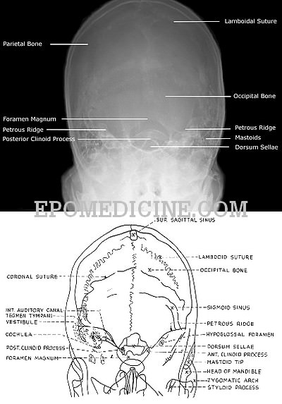
Posterior clinoid process 5. Mastoid air cells 9. X-Ray imaging for RIGHT ARM.
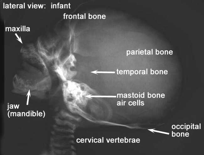
The central beam of X-rays passes from one side of the head and is at angle of 25° caudad to radiographic plate. This is an x-ray image of the skull of an infant taken from a lateral view showing the skull from the side. Head and Neck Ear Mastoiditis, Schuller view (13) AG CT MMG MRI NM RF US X-ray.
Looking for X - Ray Mastoids LAT Obl & Townes View test. Chest X-ray (Apicolordotic View) Neck X-ray;. Skull x-ray lateral view.
Radiographic Positioning of the Knee AP Views By:. We have been studying how to make x-ray examination of the temporal bone, middle ear, and mastoid process as simple and informative as possible. HRCT Stenvers reformat Stenvers plane (oblique coronal, i.e.
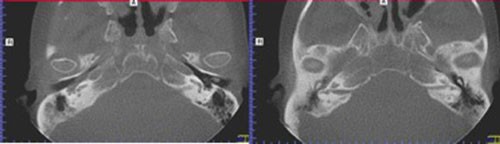
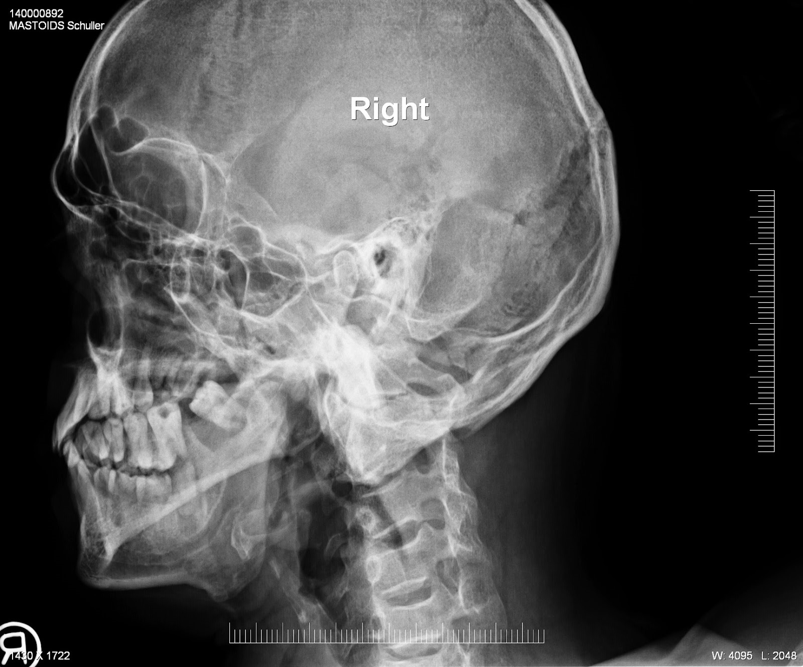
X-ray mastoids were obtained by Law's view bilaterally and high resolution computed tomography of the temporal bone was obtained with 1mm cuts in axial and coronal planes. For more information or to schedule an appointment, please call 310-423-8000. Lateral oblique (Schuller) Done when cortical mastoidectomy is required in ear discharge refractory to antibiotics.
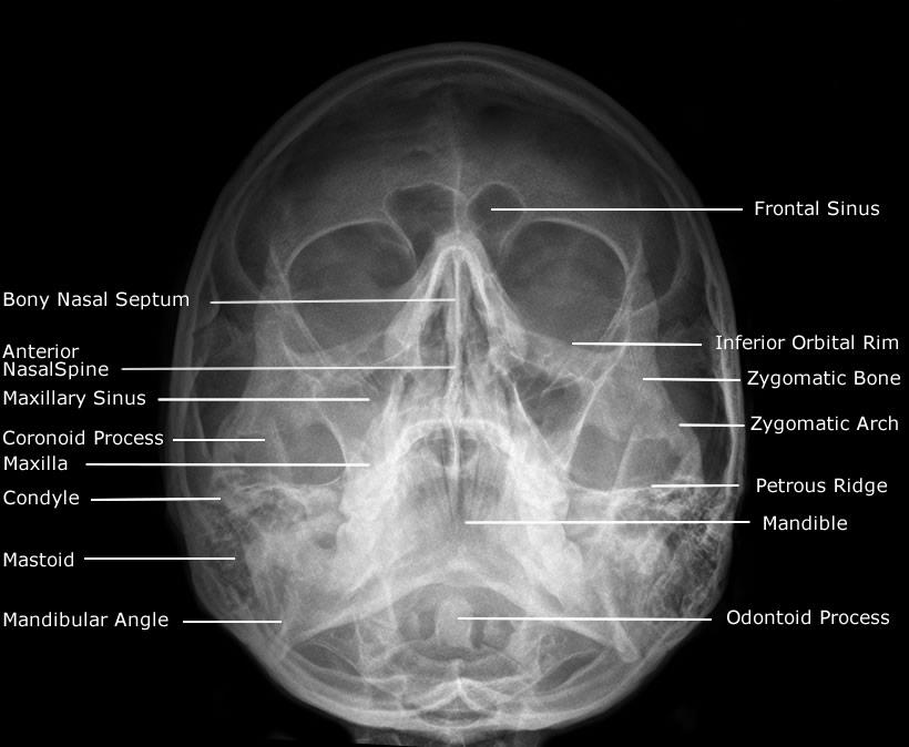
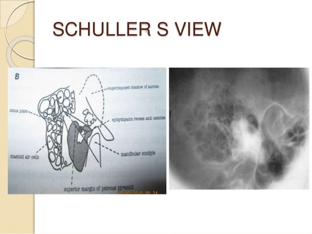
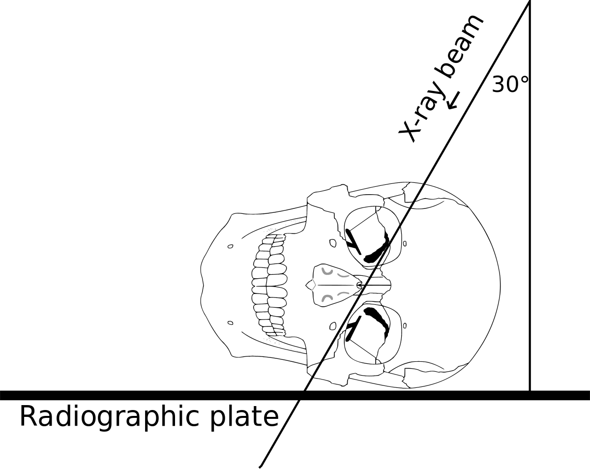
Central Ray The horizontal central ray is centered in the midline of the occiput so that the emergent ray exits the patient in the midline at the level of the anterior nasal spine at the upper border of the maxilla. 7 matched, X RAY MASTOID LEFT LAT OBLIQUE scan in (near) HARI NAGAR, NEW DELHI, Book online at HealthDx.in, compare the cost (rate) of services offererd, book your scan now!. Schüller's view (Runstrom) is a lateral view of the mastoid obtained with the sagittal plane of the skull parallel to the film and with a 30° cephalocaudal angulation of the x-ray beam.
CORRELATION OF RADIOLOGICAL AND OPERATIVE FINDING REGARDING THE CELLULARITY OF MASTOID IN CHRONIC SUPPURATIVE OTITIS MEDIA (ATTICOANTRAL DISEASE). Mark Taper Foundation Imaging Center provides a full range of advanced imaging, both radiology and cardiology, as well as interventional radiology and interventional tumor (oncology) treatments to the greater Los Angeles area, including Beverly Hills, Encino, Mid-Cities, Sherman Oaks, Silver Lake, Studio City. It is thought that CSOM is usually associated with sclerosis of the mastoid, but various authors in the past while operating on atticoantral disease ear found that the mastoid air cell.
The X-ray beam is directed either postero anteriorly or antero posteriorly along the orbito-meatal line at an angle of 90 degrees to the film. Various views for mastoid • LAW’s view- lateral Oblique view. Procedure for X-Ray Mastoid (Right) (AP View) Test Examination of the mastoid can be made possible with the AP view which is also called posteroanterior and anteroposterior.
X-ray of mastoids, frontal view. 12 degree cephalad angulation, with head rotated 45 degrees from AP. Version 2.68 -0XR Mastoid - bilateral Law and Mayer and Stenver and TowneActive Fully-Specified Name Component Views Law + Mayer + Stenver + Towne Property Find Time Pt System Head>Mastoid.bilateral Scale Doc Method XR Additional Names Short Name XR Mastoid-Bl Law+Mayer+Stenver+Towne Basic Attributes Class RAD Type Clinical First Released Version 2.14 Last Updated Version 2.64 Change.
The standard projections for the radiographic examination of mastoid include:. Infant skull x-ray lateral view. Like a usual X-ray test, low radio waves are passed through the ear and lower head portion.
Mastoid cavity & E.A.C. Skull, lateral X-ray, child 5 months. Law’s view (15º lateral oblique):.
What’s up guys, kaise ho dosto?. Anterior border of head positioner 4. Imaging the brain’s structure and examining its physiology, both in the acute and elective setting, are now the domain of multiplanar, comp.
Mastoid - Lateral Oblique. Stenvers projection taken to demonstrate internal auditory canal and temporal bones.anterior projections. 1 article features images from this case.
We make a new Technique Of mastoid Towne's View X-Ray. In most cases, a sinus X-ray will be one test performed in a series of tests. RadTechOnDuty is an Educational Blog for Technicians.
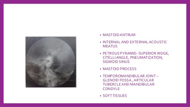
Mastoid air cells 2. Routine haematological and biochemistry investigations were done to assess fitness for anaesthesia. Schuler described the first view to visualise pathologic lesions in the area frequently involved in chronic disease namely attic, aditus, antrum or the key area.
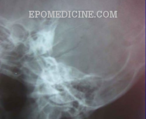
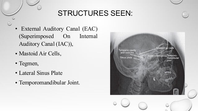
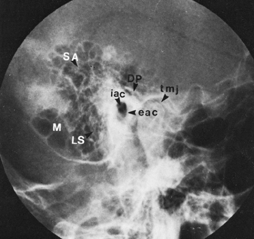
Schuller’s or Rugnstrom view (30º lateral oblique):. X-ray of mastoids, lateral and frontal views. The X-ray mastoid is done to know mastoid pneumatisation and the level of sinus and dural plates.
The X-ray beam is directed at a 15 degree oblique plain cephalocaudally while the skull's sagittal. In radiology, oblique view of skull, to establish better views for petrous bone, bony labyrinth and internal auditory canal. Radiograph for each mastoid is taken separately.
Lateral oblique (Schuller) Done when cortical PPT. Radiology Schools Radiology Student Medical Radiography Advanced Nursing Radiologic Technology Nursing Information Rad Tech Emergency Medicine Medical Imaging. A recent search of the old X-ray records and films at the Middlesex Hospital show that in September, 1946, and in January, 1947, this five-view technique was still in vogue, but that by June, 1947, a sixth view was jidded to our routine mastoid technique.
It is also called an Axio-anterior oblique posterior view. Second major axis-property observed (e.g., mass vs. Please choose Location and other options on this page to view final cost in Delhi NCR.
And Jan Žižka2 (1) Department of Radiology, University Hospital Hradec Králové, Hradec Králové, Czech Republic (2) Department of Radiology Faculty of Medicine in Hradec Králové, University Hospital Hradec Králové Charles University in Prague, Hradec Králové, Czech Republic S00:. Within 24 Hours* Test Price:. X-ray of mastoids, oblique and frontal views.
Third major axis-timing of the measurement (e.g., point in time vs 24 hours) System:. Please note that these scans involve X-Ray radiation, and are not to be performed during pregnancy. Investigations Examination under microscope C/S from discharge Rigid oto endoscopy – to see facial recess and sinus tympani if possible PTA X-ray mastoid schuller’s & laws view HRCT temporal bone 17.
Become a single cavity seperated by middle PPT. Interparietal bone (Inca bone) Anterior fontanelle;. Mastoid cavity & E.A.C.
Sagittal plane of the skull is parallel to the film and X-ray beam is projected 15. Accordingly, examination of the mastoid can be possible using the following projections:. What is required of us by the otologic surgeon is a demonstration of the middle ear and ossicles, the epitympanic space, bony bridge, aditus, and the mastoid antrum.
Sinus X-rays are less invasive than other types of sinus tests, but they’re also less comprehensive. Long axis of the pyramid) Using an MPR 3D…. The X-ray beam is directed at a 15 degree oblique plain cephalocaudally while the skull's sagittal plane is parallel to the X-ray film.
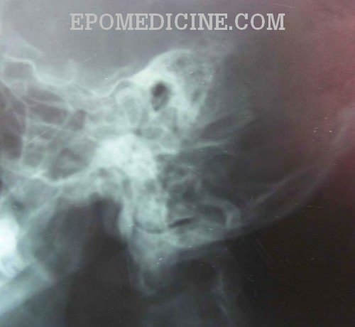
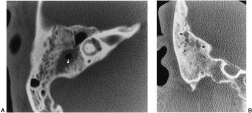
CT has typically overtaken x-ray as the modality of choice for imaging of the mastoid. First major axis-component or analyte Property:. An x-ray mastoids lateral oblique view (Laws) showed (L) mastoid to be sclerotic with evidence of bone destruction.
The oblique view X-ray test scans the mastoid bone from a lateral angle. If you have been having headaches, and ct scans have all bee. Dosto aj app dekhenge ki mastoid ka stenvers view ko kaoise kiya ja sakta hai/ dosto aj k is developed samai main s.
No Special Preparation Required Reporting :. Rarefied (almost non-existent on the left side) mastoid cells on both sides with sclerotic septa imply chronic mastoiditis. Modified Law method is an x-ray special projection to best demonstrate the abnormal relationship of temporo-mandibular fossa or TMJ, which also known as rang of motion between condyles and TM fossa.
This angulation prevents overlap of images of two mastoid bones. The X-ray beam is directed at a 14 degree angle caudally and the head faces the film with slight flexion and rotation at an angle of 45 degress to the opposite side. If you like this video Please.
Anterior clinoid process 6. VIEWS LAW + MAYER + STENVER + TOWNE:. Nasal Bone – apl (Water’s View And Soft Tissue Lat) Chest X-ray (Ap, Lat.
It has succeeded the Stenvers view, which includes more of the mastoid air cells. Download mfine app, Upload reports and Consult Top Doctors Online the minute you need to. After the examination and appropriate investigations informed consent was taken for participation in the trial.
X-ray of mastoids, oblique view. Mastoid process (Processus mastoideus) The skull is composed of multiple small bones held together by fibrous joints. Old) Nasal Bone Soft Tissue Lat;.
Mastoid X-ray (Laws & Mayers) Chest Bucky (Ap) Mastoiditis;. Performed on a Digital X-Ray. CE4RT This is an article for Radiologic Technologists (X-Ray Techs) about radiographic positioning of the knee in AP projections.
Skull X-Ray Lateral View. Similar to Law’s view but cephalocaudal beam makes. The more electrodes in the cochlea the better.
Its inferior surface gives rise to a number of projections, and these allow for the attachment of many structures of the neck and face.The temporal bone is one of the bones of the skull. I done CT brain and X-ray for mastoid and its ok." Answered by Dr. This is an X-Ray image of the Skull taken from a Lateral View showing the Skull From the Side.
Commonly this projectjion is taken in open and closed mouth position.

Mandible Flashcards Quizlet

Skeletal Identification By Radiographic Comparison Of The Cervicothoracic Region On Chest Radiographs Sciencedirect
Journals Sagepub Com Doi Pdf 10 1177
X Ray Of Mastoids Epomedicine

Pdf The Growth Rate And Size Of The Mastoid Air Cell System And Mastoid Bone A Review And Reference
Medical Imaging And Radiological Anatomy X Ray Ct Mri Kenhub
Www Jemds Com Latest Articles Php At Id 7457
X Ray Of Mastoids Epomedicine
Www Thieme Connect De Products Ebooks Pdf 10 1055 B 0034 619 Pdf

Bioactive Glass Granules For Mastoid And Epitympanic Surgical Obliteration Ct And Mri Appearance Semantic Scholar

The Use Of Radiology In Mass Fatality Events Sciencedirect
Hesi Flashcards Easy Notecards

Mastoids Radiographic Anatomy Wikiradiography Medical Radiography Radiologic Technology Radiology Student

Mastoid Series Normal Radiology Case Radiopaedia Org
Q Tbn 3aand9gcr3aalgrguwrjrisbfvpj6n60uirruhe5fghdtqrsv3urz2xkto Usqp Cau

Modified Law Method Tmj Radtechonduty
X Ray Of Mastoids Epomedicine
Http Www Neurosurgeryresident Net D diagnostics D45 59 neuroimaging X Ray ct mri pet mrs D47 x Ray Pdf
Radiology In Head And Neck By Kanato T Assumi

Petrified Ear Karthikeyan P Bala A G Priya K Indian J Otol
Www Jemds Com Latest Articles Php At Id 7457
X Ray Of Mastoids Epomedicine
Skull Radiography Techniques And Reporting
Journals Sagepub Com Doi Pdf 10 1177
The Middle Ear And Mastoid Radiology Key

Osce Notes In Otoradiology By Drtbalu Osce Notes In Otolaryngology

Ce4rt X Ray Positioning Of The Mastoid Process For Radiologic Techs
Schuller S View Wikipedia

Comparative Medical Radiography Practice And Validation Sciencedirect

Combined Use Of Frontal Sinus And Nasal Septum Patterns As An Aid In Forensics A Digital Radiographic Study

Jaypeedigital Ebook Reader
Clinical Discussion Ear Ppt Video Online Download
Roentgenology Of Skull
Pubs Rsna Org Doi Pdf 10 1148 80 2 255

Role Of X Rays In Otolaryngolgoy Pdf Document
Q Tbn 3aand9gctsnvj4ggfuru57jutyuoc5zay0w4qwkymux4wwlsfd2oyatxxv Usqp Cau
Www Jemds Com Latest Articles Php At Id 7457

Jaypeedigital Ebook Reader

Jaypeedigital Ebook Reader
Surgical Approach For Complete Cochlear Coverage In Eas Patients After Residual Hearing Loss
Medical Imaging And Radiological Anatomy X Ray Ct Mri Kenhub
Mastoid Process Anatomy Function And Attachments Kenhub

Skull Towne View Radiology Reference Article Radiopaedia Org

Case 21 1991 A 13 Year Old Boy With A Destructive Lesion Of The Left Mastoid Bone Nejm
2

Skull Caldwell Radiology Imaging Medical Radiography Radiology Student
X Ray Of Mastoids Epomedicine
The Temporal Bone Radiology Key
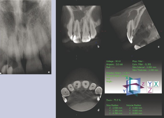
Cone Beam Ct In Dental Practice British Dental Journal
Journals Sagepub Com Doi Pdf 10 1177

Diseases Of Ear Nose And Throat 6th Edition Pages 451 491 Flip Pdf Download Fliphtml5
Presentation1 Pptx Radiological Anatomy Of The Petrous Bone
Digital X Ray Of Mastoid Region Law S Lateral Oblique View Showing Download Scientific Diagram

Conebeam Ct Of The Head And Neck Part 2 Clinical Applications American Journal Of Neuroradiology

Ce4rt X Ray Positioning Of The Mastoid Process For Radiologic Techs
Www Thieme Connect De Products Ebooks Pdf 10 1055 B 0034 619 Pdf
Pubs Rsna Org Doi Pdf 10 1148 80 2 255

Jaypeedigital Ebook Reader
Dr Sujan Chhetri Ms Ent Ppt Video Online Download

Skull Radiographic Anatomy Wikiradiography Radiology Imaging Medical Radiography Radiology Schools

Mastoids Lat Obl View Anatomy And Physiology Part 23 Youtube

Radt 086 Federal Organizations Governing Radiation Safety Regulations Youtube
Pps Radiology

Jaypeedigital Ebook Reader

Bioactive Glass Granules For Mastoid And Epitympanic Surgical Obliteration Ct And Mri Appearance Semantic Scholar

Role Of X Rays In Otolaryngolgoy Esophagus Medical Imaging
Q Tbn 3aand9gcsgezhtgnrhnt2whhbucwmirrxbxcb3iofbxhusoedpbvyd4vco Usqp Cau
Journal Of Korean Neurosurgical Society

Mastoid Series Normal Radiology Case Radiopaedia Org

Skull Fracture Www Forensicmed Co Uk

Indian Journal Of Otology Table Of Contents

Severe Craniofacial Trauma After Multiple Pistol Shots In Open Medicine Volume 14 Issue 1 19

Nose Pns Imaging Otorhinolaryngology Portal

Jaypeedigital Ebook Reader
Www Academicradiology Org Article S1076 6332 16 5 Pdf
Http Www Neurosurgeryresident Net D diagnostics D45 59 neuroimaging X Ray ct mri pet mrs D47 x Ray Pdf
Calcinosis Cutis X Ray Radiology
Flchirocon Com Wp Content Uploads 17 11 Diagnostic Imaging In The Busy Chiropratic Practice Flchirocon17 Jacksonville Final For Notes Pdf
X Ray Of Mastoids Epomedicine
Flchirocon Com Wp Content Uploads 17 11 Diagnostic Imaging In The Busy Chiropratic Practice Flchirocon17 Jacksonville Final For Notes Pdf
Radiological Imaging In Head And Neck And Relevant Anatomy
Www Thieme Connect De Products Ebooks Pdf 10 1055 B 0034 619 Pdf

X Ray Anatomy Labeling Skull Flashcards Quizlet
Pubs Rsna Org Doi Pdf 10 1148 80 2 255

Radiology In The Study Of Bone Physiology Academic Radiology

X Rays In Ent
Top Photos In Infant Skull X Ray Lateral View
Q Tbn 3aand9gcq8 5nnwyclztdh8hoevszmty72chgss1tcrgum0bew3hkaveri Usqp Cau
Nanopdf Com Download File 3159 Pdf

Mastoiditis Receiving
Journals Sagepub Com Doi Pdf 10 1177
The Temporal Bone Radiology Key

Digital X Ray Of Mastoid Region Law S Lateral Oblique View Showing Download Scientific Diagram
Pubs Rsna Org Doi Pdf 10 1148 80 2 255

500 Best Xray Critique Images In Radiology Radiography Radiology Technologist

Conebeam Ct Of The Head And Neck Part 2 Clinical Applications American Journal Of Neuroradiology

A And B X Rays Both Mastoids Law S View Showing Radio Opaque Foreign Download Scientific Diagram

The Temporal Bone Radiology Key


